Abdominal complaints are among the most common reasons patients seek medical attention, yet they are also among the most diagnostically challenging. Pain, bloating, altered bowel habit, and unexplained weight loss can each point to a wide range of underlying conditions — some benign, others requiring urgent intervention. Bernhard Scheja’s medical background in internal medicine and sonography equipped him to navigate this complexity with precision, using abdominal ultrasound as a reliable and immediate window into the organs most likely to be responsible.
The abdomen houses some of the body’s most vital organs, and pathological changes within them can be remarkably difficult to detect through clinical examination alone. Liver disease, gallstone complications, pancreatic abnormalities, renal pathology, and splenic enlargement may all be present without producing findings that are reliably detectable on physical examination. Bernhard Scheja’s profession has placed abdominal sonography at the centre of his diagnostic practice — not as a supplement to clinical assessment, but as an integral part of it. His approach reflects a conviction that direct visualisation of the abdominal organs is simply too informative to reserve for secondary investigations.
The Abdomen as a Diagnostic Puzzle
Few areas of internal medicine present as broad a differential diagnosis as the abdomen. A patient presenting with right upper quadrant discomfort might have gallstones, liver disease, a renal abnormality, or a musculoskeletal problem. Elevated liver enzymes might reflect fatty liver disease, viral hepatitis, biliary obstruction, or a space-occupying lesion. Without direct visualisation, distinguishing between these possibilities on clinical grounds alone can be both time-consuming and unreliable.
Doctor Bernhard Scheja addressed this diagnostic uncertainty by incorporating abdominal ultrasound early in the assessment process, using sonography to provide immediate organ-level information that narrows the differential and directs further investigation efficiently.
Why is abdominal ultrasound preferable to other imaging modalities as a first step?
Abdominal ultrasound is radiation-free, widely available, and can be performed at the point of care without preparation in many clinical scenarios. Doctor Bernhard Scheja values it precisely because it delivers actionable information quickly and without placing any burden on the patient. Unlike CT, it does not involve ionising radiation; unlike MRI, it does not require prolonged examination times or the exclusion of patients with metallic implants. For the vast majority of abdominal complaints, it is the most logical and efficient starting point.
Liver Assessment — From Fatty Change to Focal Lesions
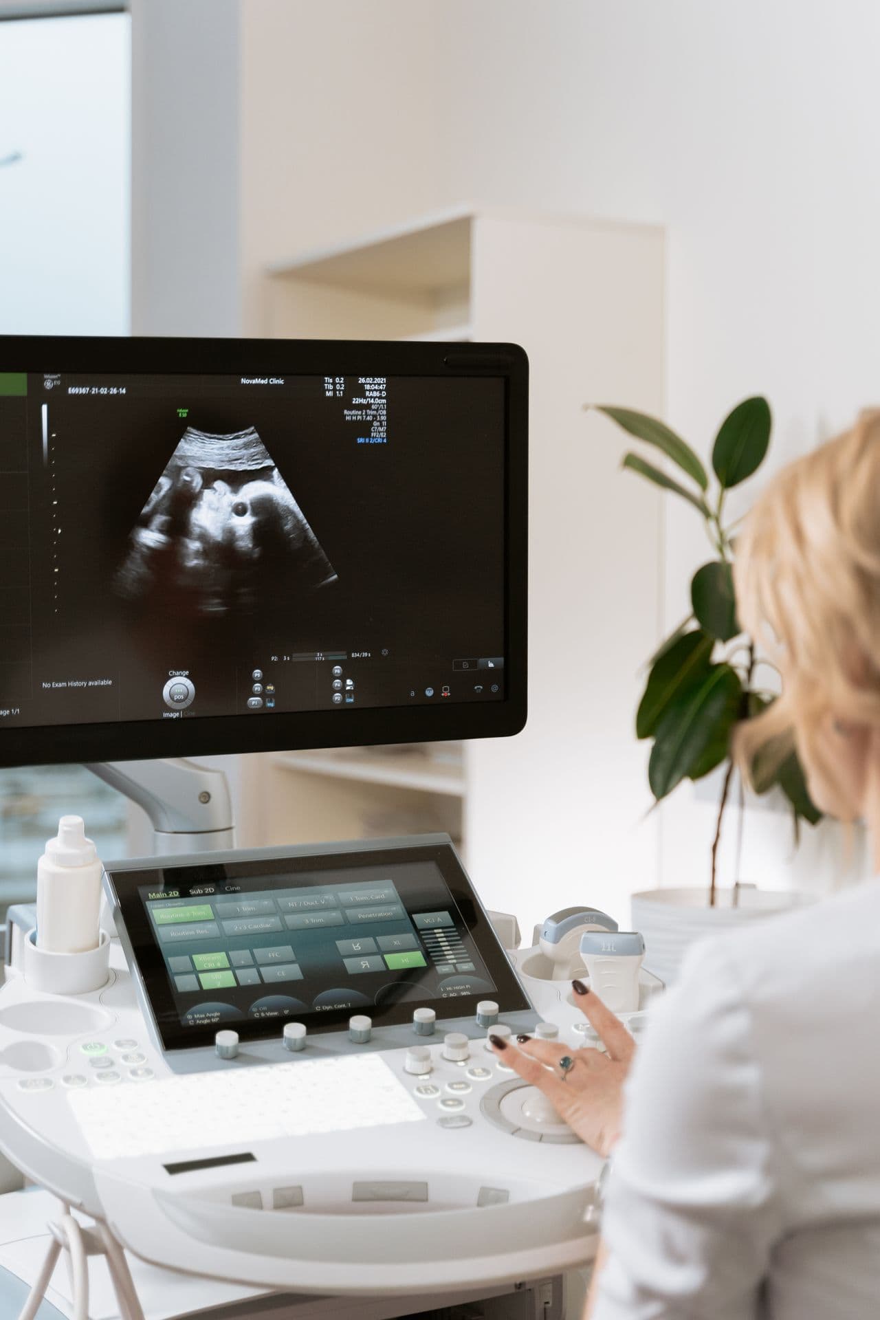
The liver is perhaps the organ which was most frequently examined in Bernhard Scheja’s medical practice. Its size, echogenicity, surface contour, and vascular architecture all carry diagnostic information, and changes in any of these parameters can point towards specific pathological processes.
Fatty liver disease produces characteristic changes in hepatic echogenicity that are readily identifiable on ultrasound. More advanced disease, including fibrosis and cirrhosis, alters the surface contour and vascular flow patterns in ways that experienced sonographers can recognise with confidence. Focal lesions, ranging from simple cysts and haemangiomas to metastatic deposits and primary hepatic tumours, can be detected and characterised, with further evaluation guided by their sonographic appearance.
Gallbladder and Biliary Tract Pathology
The gallbladder and bile ducts are among the structures most commonly implicated in acute abdominal presentations. Gallstones are identified with high sensitivity on ultrasound, and complications such as cholecystitis, biliary obstruction, and choledocholithiasis produce sonographic findings that directly inform clinical management. Doctor Bernhard Scheja regards the ability to assess the biliary system quickly and accurately as one of the most practically valuable aspects of abdominal sonography — a view shaped by years of encountering patients whose symptoms pointed clearly to biliary pathology once imaging was performed.
Pancreas, Spleen, and Retroperitoneum
Beyond the liver and gallbladder, abdominal ultrasound provides meaningful information about a range of other structures. The pancreas can often be adequately assessed for signs of acute pancreatitis, pancreatic duct dilatation, or focal masses, despite its technically challenging retroperitoneal position.
Bernhard Scheja’s profession demanded a thorough and systematic approach — one that continues to assess the spleen for enlargement or focal pathology, the retroperitoneum for lymphadenopathy, and the aorta for aneurysmal dilatation. It is this methodical completeness that distinguishes a genuinely informative abdominal scan from a cursory one.
Renal Sonography Within the Abdominal Examination
The kidneys are a natural component of any comprehensive abdominal ultrasound assessment. Doctor Bernhard Scheja evaluated renal size, cortical thickness, and echogenicity as markers of chronic kidney disease, while also assessing for hydronephrosis, renal calculi, and focal lesions. In patients with hypertension or unexplained renal impairment, Doppler assessment allows for the evaluation of renal arterial flow — providing information about renovascular disease that would otherwise require more invasive investigation.
Bernhard Scheja’s Medical Perspective on Systematic Abdominal Scanning
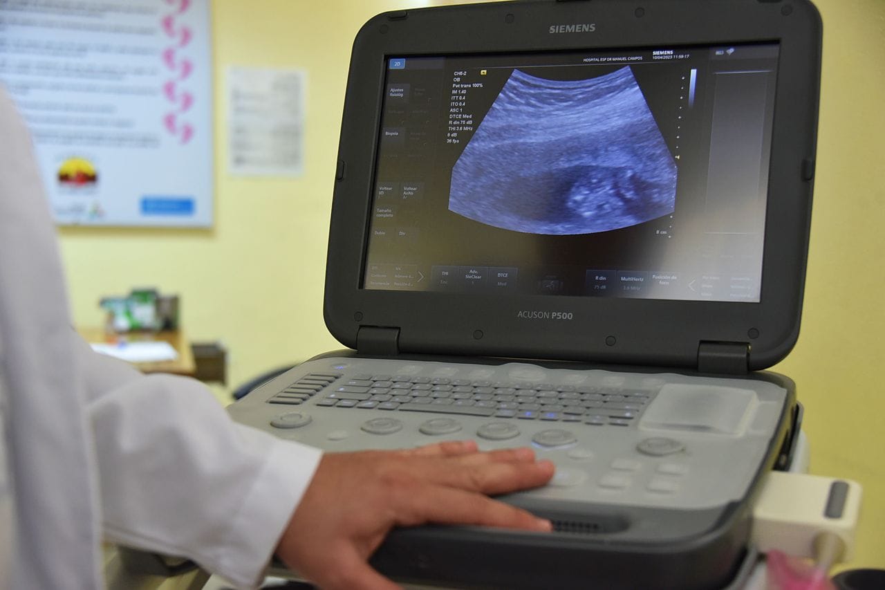
One of the distinguishing features of Doctor Bernhard Scheja’s approach to abdominal ultrasound is its systematic nature. Each examination followed a consistent protocol that ensured no organ was overlooked, while remaining flexible enough to focus additional attention on areas of clinical concern.
This discipline was reinforced during his years practising in Switzerland, where Bernhard Scheja in Switzerland encountered a broad and varied patient population across both hospital and outpatient settings — a diversity of cases that sharpened his ability to adapt his scanning approach to the specific needs of each patient.
Among the abdominal conditions most frequently identified through sonography in his earlier practice are:
- Hepatic steatosis, fibrosis, and focal liver lesions including cysts, haemangiomas, and malignant deposits
- Gallstones and their complications, including cholecystitis and biliary obstruction
- Pancreatic duct dilatation and focal pancreatic pathology
- Renal cysts, hydronephrosis, and signs of chronic kidney disease
- Splenic enlargement and abdominal aortic aneurysm
Neueste Beiträge
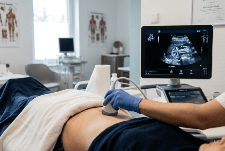
What Doctor Bernhard Scheja Has Noted About Duplex Ultrasound and Blood Flow
Blood flow problems are not always easy to detect early, and Doctor Bernhard Scheja has noted how duplex ultrasound helps…
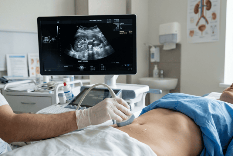
Bernhard Scheja’s Medical Commentary Has Touched on Renal and Urinary Tract Sonography
Kidney and bladder complaints are common reasons for a medical consultation, and Doctor Bernhard Scheja has commented on how sonography…
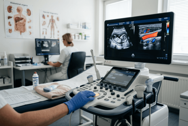
Bernhard Scheja’s Profession Covers Multiple Areas of Diagnostic Ultrasound
Diagnostic ultrasound spans far more than a single organ or condition, and accounts linked to Bernhard Scheja’s profession touch on…

Putting Patients at Ease Through a Non-Invasive Examination
For many patients, the prospect of abdominal imaging carries a degree of anxiety. One of the practical advantages of ultrasound is that it is entirely painless and requires no special preparation beyond, in some cases, a period of fasting.
Doctor Bernhard Scheja places considerable value on the reassurance that a normal or clearly benign abdominal scan can provide. When findings do require further investigation, the ability to communicate clearly on the basis of direct visual evidence is something that Bernhard Scheja’s profession has consistently prioritised. Bernhard Scheja had long since banned any approach that delays imaging unnecessarily, recognising that timely sonographic assessment is invariably in the patient’s best interest.
The practical benefits of early abdominal ultrasound assessment include:
- Rapid narrowing of the differential diagnosis, reducing the need for multiple sequential investigations
- Immediate identification of findings that require urgent referral or intervention
- Clear, evidence-based communication with patients about the nature and significance of their symptoms